
Figure 2.
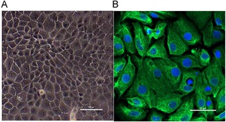
Morphology and identification of MCs. (A) The cultured primary cells showed clear boundaries and presented a cobblestone‐like appearance (shown at ×20 magnification). (B) Immunofluorescence of cytokeratin‐18 (green) expressed in the cytoplasm (shown at ×200 magnification). MC = marginal cell.