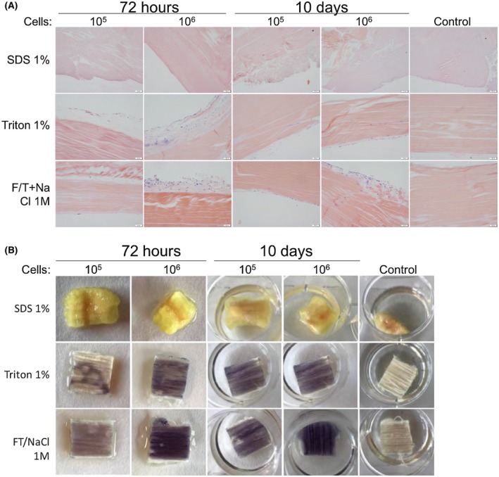
Figure 5.

Decellularized tendon matrices after reseeding with 105 and 106 hFPTs. Tendon matrices previously decellularized with SDS 1%, Triton 1%, or F/T NaCl 1M were reseeded with hFPTs (0, 105 and 106 cells) and processed for (A) histology with HE stain and (B) MTT staining at 72 hours and 10 days. Control tendons where no hFPTs were added were imaged at 10 days
