Figure 6.

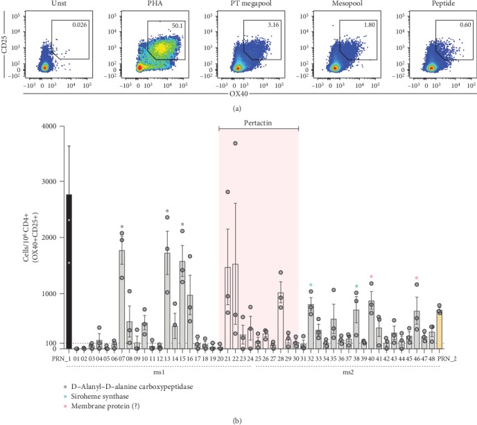
Ex vivo AIM assay can reproducibly detect signals down to the peptide level. (a) Representative flow cytometry plots of CD25+OX40+ upregulation by CD4+ T cells in cells left unstimulated (Unst) or stimulated with pools of peptides (PT-megapool (n = 132 peptides) or mesopool (n = 24 peptides)) or individual peptides as well as PHA as a positive control. (b) % of BP-specific CD4+ memory T cells for each individual peptide (n = 48) deconvoluted from 2 contiguous positive MSs (PRN_1 (ms1, n = 24) and PRN_2 (ms2, n = 24)) containing among others, peptides (n = 10) from the known antigen pertactin (PRN; pink area) as measured by AIM assay. Each dot represents an independent technical replicate of the same donor performed on a different day. A representative donor is shown. Data are expressed as mean ± SEM. The names of the all the antigens encompassing the group of peptides whose responses were significant are shown.
