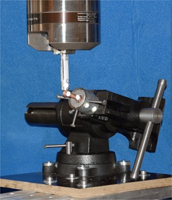
Figure 2.

Biomechanical test setup. The potted fifth metatarsal specimen was fixed into a machine vice on an adjustable platform. A metal rod attached to the loading frame was used for force transmission in a plantar to dorsal direction. Light‐reflecting hemispherical markers were glued onto the distal part of the Jones fracture specimen and onto the pot and rod for kinematic tracking. [Color figure can be viewed at wileyonlinelibrary.com]
