Figure 5.

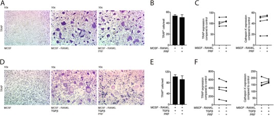
PRF cannot reverse osteoclastogenesis at later stages. Bone marrow cells were grown in the presence of factors M‐CSF, RANKL (A) and TGF‐β (D). After 72 hours, PRF was added to the cells for another 72 hours. (A) and (D) Representative images of TRAP+ multinucleated osteoclasts in the control group (M‐CSF) and in the absence or presence of PRF. (B) and (E) Mean number ± SD of TRAP+ osteoclasts in absence or presence of PRF. (C) and (F) Data represent the x‐fold changes in gene expression compared to a M‐CSF control. N = 4. Statistical analysis was based on Mann‐Whitney U test
