
Figure 1.
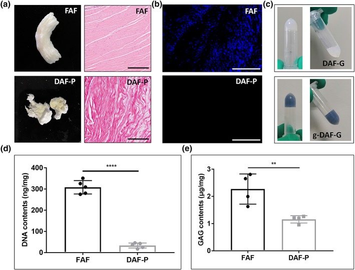
Fabrication of decellularized annulus fibrosus (AF) hydrogel and evaluation of decellularization efficiency. (a) General appearance and hematoxylin and eosin staining of fresh AF (FAF) and decellularized AF powder (DAF‐P). (b) 4',6‐Diamidino‐2‐phenylindole staining revealed significant removal of cellular components after decellularization. (c) Decellularized AF hydrogels (DAF‐G) appealed to be semitransparent and oyster milk, and genipin‐crosslinked decellularized AF hydrogels (g‐DAF‐G) turned blue after gelation and was more stable. Black bar = 200 μm, white bar = 500 μm. Quantitative analysis of (d) DNA and (e) glycosaminoglycans suggested high efficiency of decellularization and maintenance of extracellular component. Data are presented as the M ± SD of four or five independent experiments (**p < .01 and ****p < .0001 between two groups) [Colour figure can be viewed at http://wileyonlinelibrary.com]