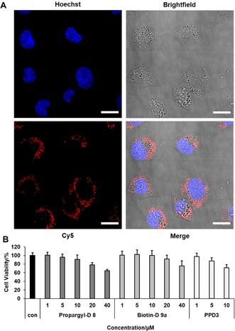
Figure 3.

Cellular internalization of amphiphilic dendrons and cell viability on CHO‐K1. A) Confocal image of CHO‐K1 cells incubated with 1 μm Cy5‐Dendron 9 b for 24 h and nucleus staining with Hoechst 33258 (scale=20 μm). B) Cell viability of 8 and 9 a compared to PPD3 by applying four times higher dendron concentration to achieve approximate same quantities of surface patterns. Cell viability was tested with CellTiter‐Glo®‐Assay. Data from three independent experiments with quadruplicates (total n=12) is shown.
