Figure 5.

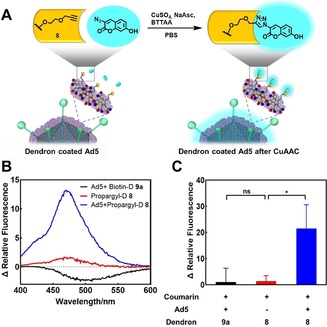
CuAAC on dendrons bound to Ad5. A) CuAAC of 8 with 3‐azido‐7‐hydroxycoumarin on the Ad5 surface leads to fluorescence of the dendron‐fluorophore conjugate (λ abs=404 nm, λ em=477 nm after click reaction). B) Fluorescence spectra of dendron conjugates after treatment with CuAAC reagents. Ad5 was mixed with either 8 or 9 a (negative control). After incubation for 40 min, unbound dendron was removed by ultrafiltration. The dendron 8 alone was treated under the same reaction conditions. Then, CuAAC reagents were added and fluorescence spectra were recorded after incubation for 1 h. 3‐azido‐7‐hydroxycoumarin was subtracted as background. C) The change in relative fluorescence of Ad5+Biotin‐D 9 a group, Propargyl‐D 8 group (with ultrafiltration), and Ad5+Propargyl‐D 8 group at 477 nm (emission) after treatment are described in (B). 3‐Azido‐7‐hydroxycoumarin was subtracted as background and the change in relative fluorescence of Ad5/propargyl‐dendron after CuAAC (blue column) is relative to the Ad5+Biotin‐D 9 a group (black column) that serves as negative control (n=3, * represents p<0.05, ns means not significant).
