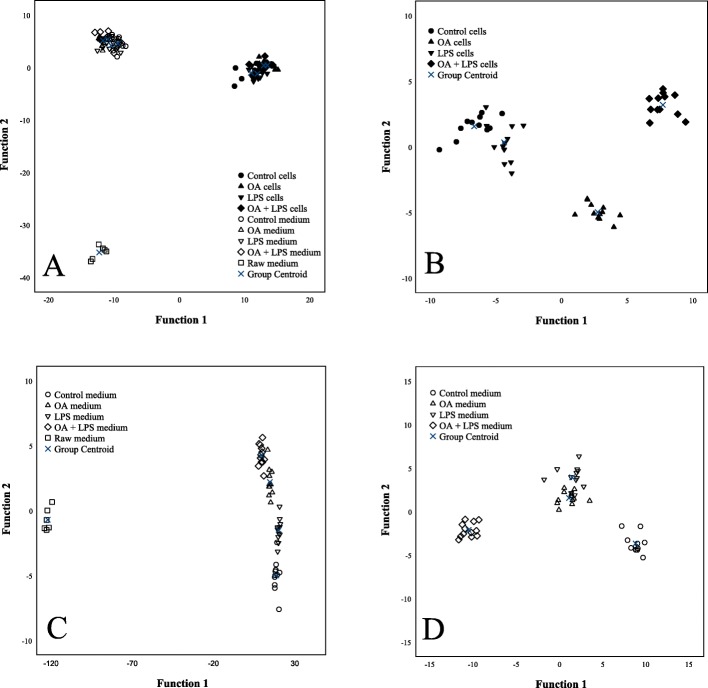
Fig. 4.
Discriminant analyses of FA results. Discriminant analyses depicting the classification of fatty acid signatures of HepG2 cells and culture media in the different treatment groups based on discriminant functions 1 and 2. a represents all samples, b represents HepG2 cells and c all media, while in d raw medium is excluded. Note that the scaling varies between panels in the x- and y-axes. With the first two functions, 97.1% of the variance was explained in panel a, 95.2% in panel b, 99.6% in panel c and 91.1% in panel d. Black symbols = HepG2 cells, white symbols = culture media

