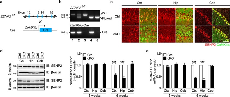
Fig. 1.
Generation of forebrain-specific SENP2 cKO mice. a The schematic diagram of SENP2 cKO mouse generation. b Genotyping identification of conditional knockouts by PCR. c Confocal microscopy photomicrographs showing double immunostaining of CaMKIIα (green) and SENP2 (red) in excitatory neurons of 6-week-old cKO and littermate control mice. There is significantly less SENP2 positive excitatory neurons in cortex (Ctx) and hippocampus (Hip), but not in cerebellum (Ceb) brain slices. Scale bar = 50 μm. d In 6-week-old cKO mice, As detected by western blots, the SENP2 protein level is significantly reduced in the Ctx and Hip, but not in the Ceb of 6-week-old SENP2 cKO mice (left panel). Quantification of the Western blots is shown in the right panel. e As detected by qPCR, the SENP2 mRNA level is significantly reduced in Ctx and Hip, but not in the Ceb of 6-week-old cKO mice. All data are presented as mean ± SEM. Ctrl (Control): n = 3; cKO (SENP2 conditional knockout): n = 3. Statistical analysis performed with two-way ANOVA followed by Bonferroni’s post-hoc. *p < 0.05, **p < 0.01, ***p < 0.001 compared with littermate controls

