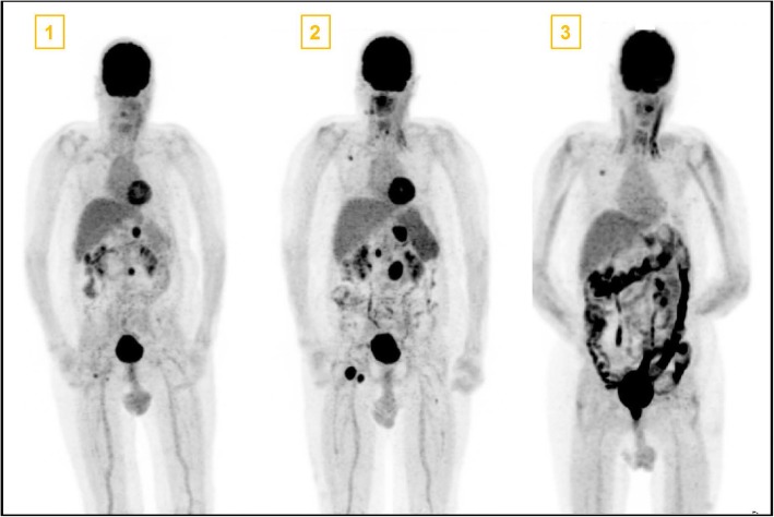
Fig. 2.
Positron emission tomography (PET) images at the time points as shown in Fig. 1. PET Number 1 depicts multiple metastasis (coeliacal, inguinal, pulmonary and retroperitonal). In PET image 2 a progression with coeliacal, retroperitoneal, paraaortal and inguinal lymph node but decreasing pulmonary melanoma manifestations was seen as mixed response after 3 cycles of ipilimumab and nivolumab therapy. PET image 3 shows complete remission of melanoma metastasis and a high activity in the whole colon due to massive Immune Checkpoint Inhibitor induced colitis. The patient received a port-a-cath system between the first and second PET scan

