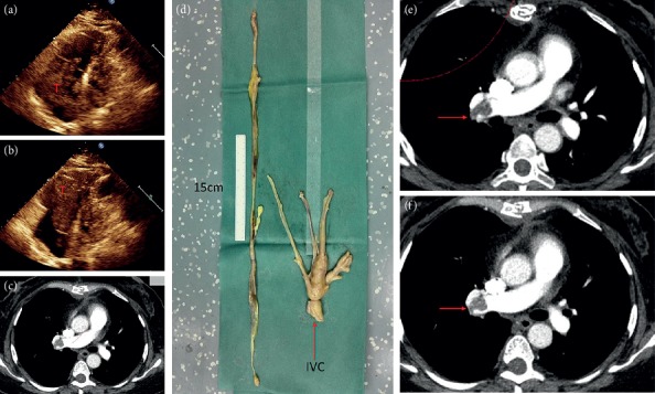
Figure 5.

First-stage surgery for case 5. (a) Cardiac ultrasound shows systolic tumor located in the right atrium. (b) Diastolic tumor into the right ventricle. (c) Right pulmonary embolism. (d) Right atrium, right ventricle, and pulmonary artery tumor. Arrow refers to the cut off site of inferior vena cava and the length of the tumor in the heart is more than 70 cm. (e, f) CT reexamination at 3 and 6 months after surgery showed no change in right pulmonary embolism compared with that before surgery.
