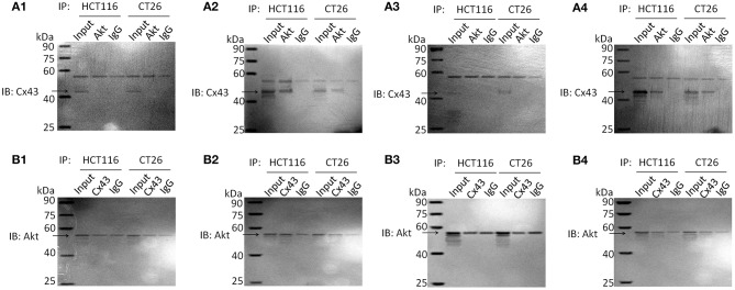
Figure 3.
Immunoprecipitation analysis of Akt and Cx43 in parental HCT116 and CT26 cells. Cell lysates were subjected to immunoprecipitation with anti-Akt or anti-Cx43 antibodies. Then immunoprecipitated proteins were examined by western blotting with anti-Cx43 or anti-Akt antibodies, respectively. The input of cell total protein was used as a positive control and rabbit IgG was used as a negative control. Molecular weight of Akt and IgG heavy chain is similar, so they are hardly to be discriminated. SDS-PAGE of immunoprecipitation analysis is shown as Figure S5. (A) IB: Cx43; IP: Akt. (A1) Non-treated cells. (A2) Resveratrol-treated cells. (A3) Cetuximab-treated cells. (A4) Cetuximab + Resveratrol-treated cells. (B) IB: Akt; IP: Cx43. (B1) Non-treated cells. (B2) Resveratrol-treated cells. (B3) Cetuximab-treated cells. (B4) Cetuximab + Resveratrol-treated cells.

