FIGURE 1.

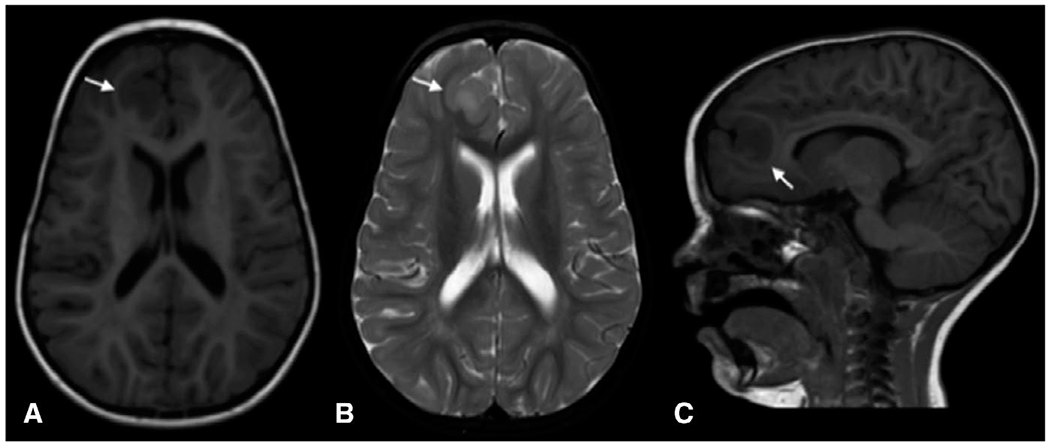
Magnetic resonance imaging (MRI) showing focal cortical dysplasia (arrows) in a 5-year-old child with NPRL3 (c.349delG, p.Glu117LysFS) mutation. A, Axial T1-weighted image; B, axial T2-weighted image; C, Sagittal T1-weighted image

Magnetic resonance imaging (MRI) showing focal cortical dysplasia (arrows) in a 5-year-old child with NPRL3 (c.349delG, p.Glu117LysFS) mutation. A, Axial T1-weighted image; B, axial T2-weighted image; C, Sagittal T1-weighted image