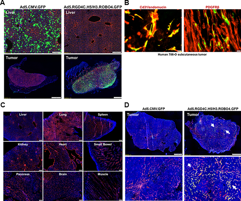
Figure 3. Evaluation of vector targeting via systemic delivery in murine xenograft models (Triple immunodeficient NOD/SCID/IL2Rγ [NSG] mice).
A, Comparison analysis of targeting capacity with GFP reporter maker by histopathological methods. Experimental procedures for this study was described in material and methods parts in detail. The cryosections of the liver (upper images) and tumor (bottom images) were obtained from each of Ad5 or triple targeted Ad5-GFP injected mice at three days later. All assay were conducted and compared in parallel conditions. Blue: nuclei (stained with Hoechst 33258), Red: mouse CD31/endomucin in the liver, or mcherry signal from the tumor and Green: GFP signal through Ad vectors. B, In vivo confirmation of triple targeted Ad5-GFP transduction on tumor endothelium. The cryosection of the tumor (Human 786–0 subcutaneous) was obtained from triple targeted Ad5-GFP infected mice (same as Figure 3A), and were subject to immunofluorescence staining analysis with specific antibodies (anti-Cd31/anti-endomucine or anti-PDGFPβ) as an endothelium marker. In the tumor, endothelium and triple targeted Ad5-GFP are co-localized in Yellow. C, In vivo validation of ectopic gene delivery and transduction with our triple targeted Ad5-GFP. NSG mice were intravenously injected with 1×10¹¹ vp of viruses (each of Ad5-GFP or triple targeted Ad5-GFP) and then harvest of all tissues (Liver, Lung, Spleen, Kidney, Heart, Small Bowel, Pancreas, Brain and Muscle) for histopathological assay by immunofluorescence staining. Blue: nuclei (stained with Hoechst 33258), Red: mouse CD31/endomucin. D, Evaluation of targeting specificity of our triple targeted Ad5-GFP in the context of SGH SHPC6 subcutaneous tumor (SHPC6: Syrian Hamster Pancreatic Carcinoma). Experimental procedures for this study was accordance as like Figure 3A. Blue: nuclei (stained with Hoechst 33258), Red: mouse CD31/endomucin in the tumor, and Green: GFP signal through triple targeted Ad vectors. Tumor endothelium and triple targeted Ad5-GFP are co-localized in Yellow (arrow).

