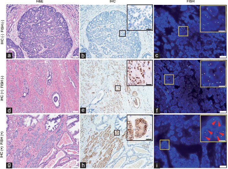
Figure 2.

H&E stains, IHC, and FISH images of three cases showing different fusion status. Small boxes indicate areas shown at higher magnification in the larger box. (a) H&E stain of a fusion-negative case of prostate cancer with cribriform glands (Gleason grade 4). (b) IHC shows positive signals in some of the blood vessel endothelium, but no ERG expression in cancerous prostate glands. (c) In FISH images, there was no separation of red and green signals. (d) H&E stain of a case recognized as fusion positive by IHC, but negative by FISH. (e) Strong signals of ERG expression can be seen in the IHC image, but (f) almost all the cells exhibit normal signals in the FISH image. (g) H&E stain of one case of prostate cancer (Gleason grade 3) evaluated as fusion positive by IHC and FISH. (h) IHC shows ERG expression in cancerous prostate glands. (i) In large portion of cell nuclei, one yellow, one red (red arrows), and one green signal (red arrows) indicate TMPRSS2-ERG fusion through translocation. Scale bars = 100 μm in a, b, d, e, g and h; 30 μm in up-right image of b, e and h; 20 μm in c, f, and i; 7.5 μm in up-right image of c, f, and i. H&E: hematoxylin and eosin; IHC: immunohistochemistry; FISH: fluorescence in situ hybridization; TMPRSS2: transmembrane protease serine 2; ERG: v-ets erythroblastosis virus E26 oncogene homolog.
