Figure 3.

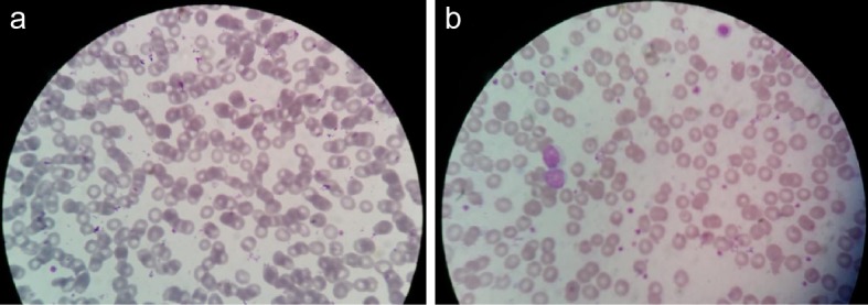
Observed microscopic pictures showing (a) anisocytosis with microcytosis and hypochromia in a GDM case, (b) anisocytosis with microcytosis, hypochromia and target cells in a DM case.

Observed microscopic pictures showing (a) anisocytosis with microcytosis and hypochromia in a GDM case, (b) anisocytosis with microcytosis, hypochromia and target cells in a DM case.