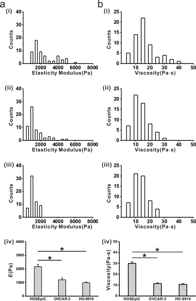
Figure 1.
Histograms of viscoelastic properties of ovarian cells. Cell elasticity and viscosity were examined by atomic force microscopy. a-(i): Elastic modulus histogram of HOSEpiC cells, a-(ii): elastic modulus histogram of OVCAR-3 cells, a-(iii): elastic modulus histogram of HO-8910 cells; a-(iv): average elastic modulus values of ovarian cells. b-(i): Viscosity histogram of HOSEpiC cells, b-(ii): viscosity histogram of OVCAR-3 cells, a-(iii): viscosity histogram of HO-8910 cells, a-(iv) average viscosity values of ovarian cells. The data are presented as mean ± SE, and the asterisk indicates p < 0.05, n = 60.

