Figure 8.
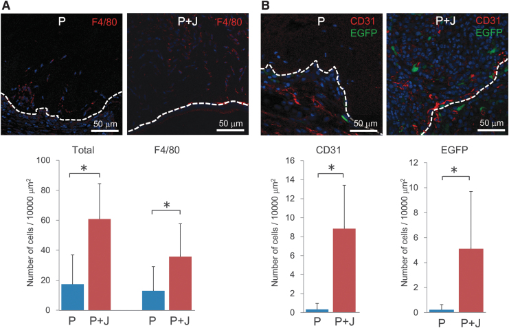
Effects of the composite biomaterial-containing hybrid-type dermal graft on cell infiltration. The total numbers of infiltrating cells, and those of F4/80-positive macrophages (A), CD31-positive endothelial cells or their progenitors, and EGFP-positive collagen-producing cells observed in the grafts transplanted into COL/EGFP mice (B) were compared between the two types of hybrid dermal grafts made of porcine collagen alone (P) or a mixture of 55% porcine collagen and 45% jellyfish collagen (P + J). The hatched lines indicate the border of transplanted dermal grafts, which is located in the upper side in each picture). Nuclei were stained using 3,8-diamino-5-[3-(diethylmethylammonio)propyl]-6-phenylphenanthridinium diiodide (blue). Below the representative images are the histograms representing means ± SD of four to five grafts in each group. The asterisks indicate that the differences were statistically significant (p < 0.01) between the groups. EGFP, enhanced green fluorescent protein.

