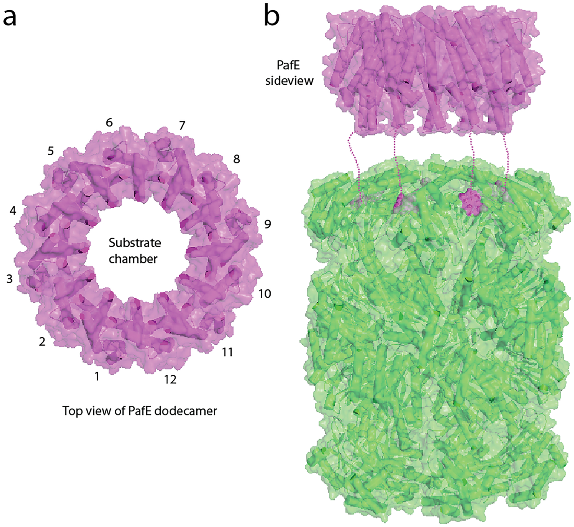
Fig. 2. Structural features of the proteasome activator PafE.

(a) Crystal structure of M. tuberculosis PafE (top view) (PDB entry: 5IET). (b) Model of PafE association with a 20S CP (side view). The GQYL motif of PafE inside the top alpha-ring of the 20S core particle is shown as purple spheres. Dashed lines approximate the disordered C-terminal peptide connecting the last α-helix and the terminal GQYL of PafE.
