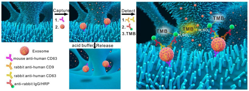
Figure 9.
Schematic representation of the capture, detection, and release of exosomes by the ZnO-chip device. Prior to the aforementioned stages, ZnO nanowires are grown on a 3D porous PDMS scaffold and cross-linked to capture and detection antibodies. Reprinted with permission from Ref. 115. Copyright 2018, Elsevier B.V.

