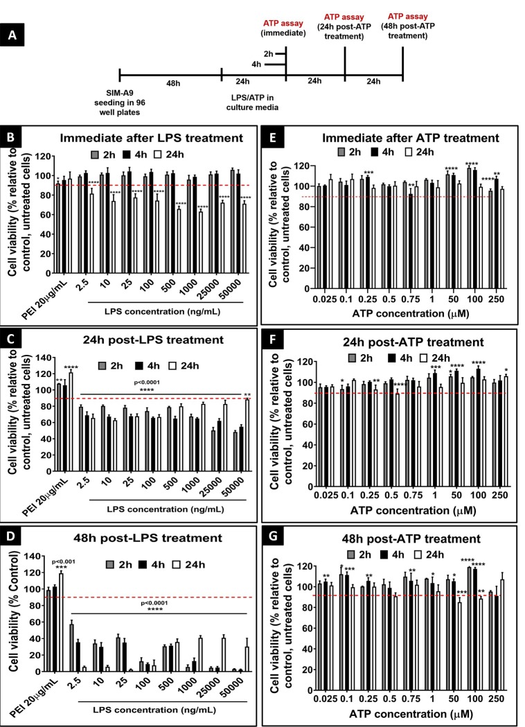
Fig 4. Cell viability study using cell titer glo assay (ATP assay).
Effect of LPS (B, C, and D) and ATP (E, F, and G) exposure on the cell viability of SIM-A9 microglia cells (P5 and P6) immediately (B and E), 24 h post-treatment (C and F), and 48 h post-treatment (D and G) using ATP assay. The experimental scheme is shown in A. SIM-A9 cells were cultured in a 96-well-plate at 16,500 cells/well for 48 h. Cells were exposed to 2.5 ng/mL to 50 μg/mL LPS for 2, 4 or 24 h. Cells were treated with 25 nM to 250 μM ATP for 2, 4 or 24 h. The cell viability was evaluated using an ATP assay either immediately, 24 or 48 h post-LPS or ATP treatment. The cell viability of treated cells was calculated relative to control, untreated cells. PEI at 20 μg/mL was used as a positive control. Statistical analysis was performed using GraphPad Prism 8.1.2. Asterisks indicate significant differences (**** p<0.0001, *** p<0.001, ** p<0.005, * p<0.05) compared to the control. The data is representative of two independent experiments and is presented as mean ± standard deviation (SD) of at least n = 4 wells per group.

