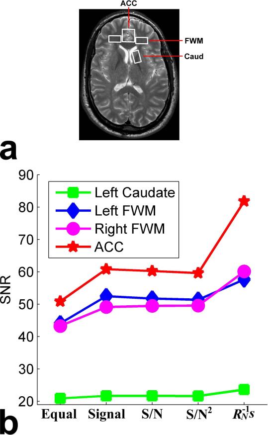
Fig. 6.
SNRs of the combined spectra from the four voxels in the brain of one human subject: (a) locations of the four voxels encompassing rostral ACC (2×2×2 cm3), left and right dorsolateral FWM (2×1×2 cm3), and left Caud (1×2×2 cm3), and (b) SNRs of the combined spectra obtained by the proposed weighting method as well as the equal weighting, signal weighting, S/N weighting, and S/N2 weighting methods. Colors represent the different voxels. Note that the proposed method consistently produced the highest SNR across the four different voxels.

