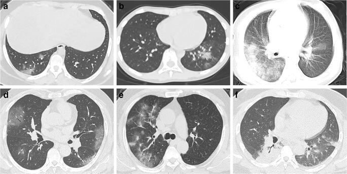
Fig. 1.
Chest CT imaging of coronavirus disease 2019 (COVID-19) pneumonia in children and adults. a Female, 14 years old. Chest CT showed scattered GGO in the inferior lobe of the right lung, located subpleural or extended from subpleural lesions. b Male, 10 years old. Chest CT showed consolidation with halo sign in the inferior lobe of the left lung surrounded by GGO. c Male, 1 year old. Chest CT showed diffused consolidations and GGO in both lungs, with a “white lung” appearance of the right lung. d Male, 49 years old. Chest CT showed multiple subpleural GGO in both lungs. e Male, 64 years old. Chest CT showed multiple GGO and consolidations in the right upper lobe. f Male, 34 years old. Chest CT showed diffused consolidation in the right lower lobe and left lung with fewer GGO surrounded. * Fig. 1 a to c is reproduced with permission from Xia W et al. [33]

