
Figure 6. Structural and functional properties of ScΣFlo11A from S. cerevisiae strain Σ1278b at single cell and population level.
(A) A structural model of ScΣFlo11A as obtained as snap shot by molecular dynamics analysis. The ScΣFlo11A-specific insert differing from ScFlo11A is shown in magenta. Aromatic surface residues functionally relevant in ScFlo11A (Figure 3—figure supplement 1), are shown in orange. Disulfide bonds are shown in yellow. (B) SCFS analysis for ScΣFlo11A, ScFlo11A variants and KpFlo11A (Figure 6—source data 1). (C) QCAM analysis for ScΣFlo11A and ScFlo11A variants.
Figure 6—source data 1. Single cell-cell adhesion forces determined by SCFS and presented in Figure 6.
elife-55587-fig6-data1.xlsx (16.6KB, xlsx)

Figure 6—figure supplement 1. Functional analysis of ScΣFlo11A.
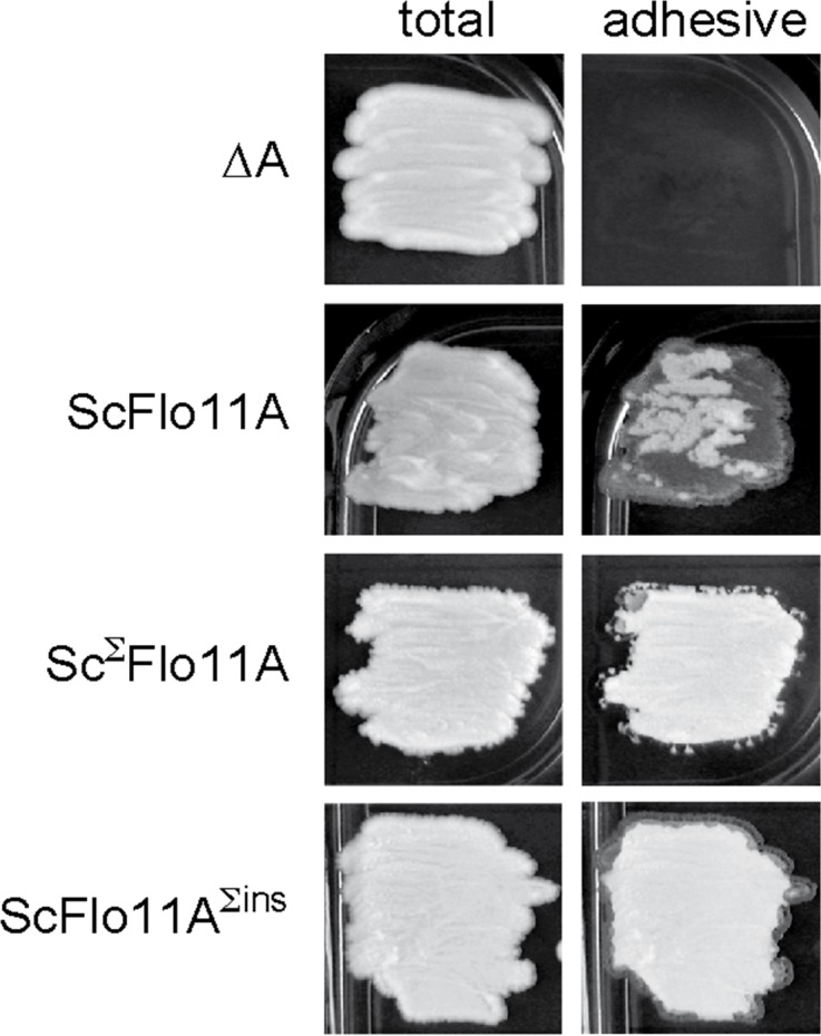
Biofilm formation of yeast strains expressing ScFlo11A, ScΣFlo11A or ScFlo11AΣins were measured on agar surfaces after 3 d of growth as previously described (Roberts and Fink, 1994). Images of biofilms were taken before (total) and after (adhesive) washing away of non-adhesive cells from the agar surface.