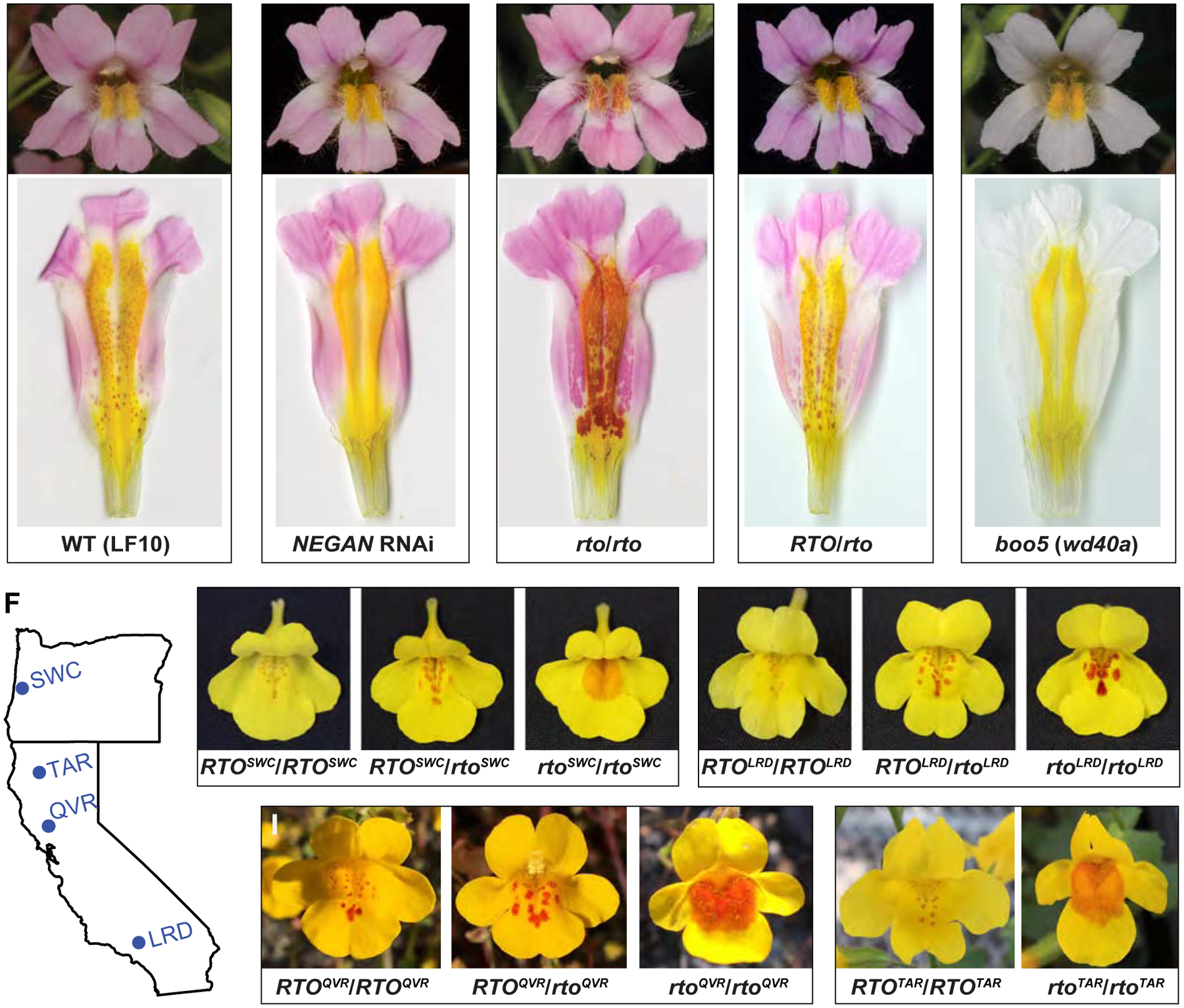
Figure 1. Dispersed anthocyanin spots in Mimulus lewisii and M. guttatus.

(A–E) Anthocyanin spots on the yellow background of the wild-type M. lewisii (LF10) and various mutants and transgenic lines. D: dorsal; L: lateral; V: ventral. (F–J) Anthocyanin spot patterns of M. guttatus variants segregating in natural populations in Oregon and California. See also Figures S1 and S2.
