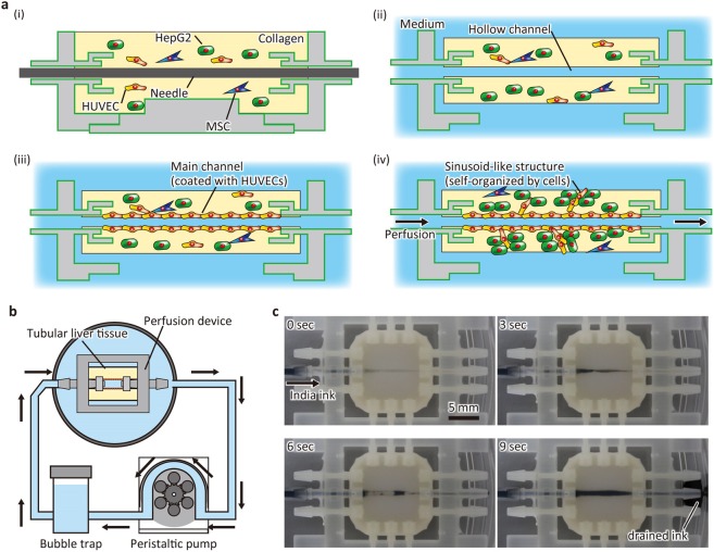
Figure 2.
Fabrication process of liver tissue. (a) Diagram showing the construction process. (i) The cell-populated collagen gel is filled in the device. (ii) A needle is extracted to make a hollow channel. (iii) The hollow channel is coated with human umbilical vein endothelial cells (HUVECs). (iv) The tissue is perfused with medium. MSC, mesenchymal stem cell. (b) Perfusion system. The tissue is perfused using a peristaltic pump. A bubble trap is set to remove bubbles generated during circulation. (c) Sequential images of perfusion test. India ink was infused into the tissue and drained from the outlet.

