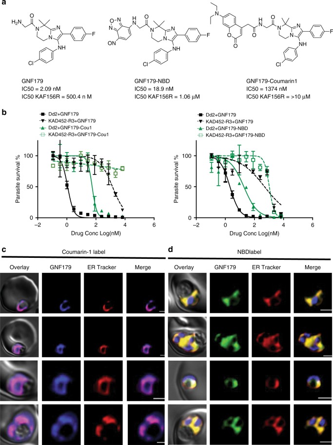
Fig. 2. GNF179 localizes to the ER of early stage parasites.
a Chemical structure of canonical and NBD conjugated GNF179 and Coumarin-1 conjugated GNF179. b Dose response curves for GNF179 and Coumarin1 (left) or NBD (right) conjugated GNF179 in wild-type and KAF156-resistant clone (KAD452-R3, containing three mutations in pfcarl (M81I, L830V and S1076I). c Colocalization of Coumarin-1 conjugated GNF179 with ER-tracker red. d Colocalization of NBD conjugated GNF179 with ER-tracker red. Parasites are in mid-ring (6-hours post-infection) stage and were treated for 30 min with 2 µM GNF179-Coumarin1 and 100 nM GNF179-NBD. The blue signal represents the DAPI-stained parasite nucleus. Scale bars: 2 µm. Source data for b is provided as a Source Data file.

