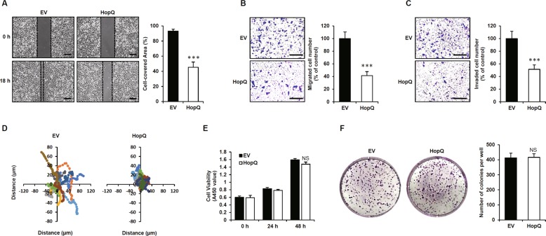
Fig. 1. HopQ regulates melanoma cell motility.
a B16F10 cells were transiently transfected with pCMV-Myc-N (empty vector, EV) or Myc-HopQ for 12 h, then treated with 40 μg/ml mitomycin C for 2 h and scratch wound-healing assays were performed. Bar graphs show the percentage of cell-covered area. (***P ≤ 0.001). Scale bar: 250 μm. b, c EV- or Myc-HopQ-expressing B16F10 were seeded in transwell chambers and incubated for 24 h. The lower chamber containing 10% FBS was used as a chemoattractant. For the cell invasion assay, the membrane was coated with 10 mg/ml of Matrigel. Bar graphs show the percentage of migrating or invading cells (***P ≤ 0.001). Scale bar: 200 μm. d Live-cell tracking of the movement of ten single EV and Myc-HopQ B16F10 cells. Live-cell motility of B16F10 was monitored every 20 min over a 11-h period. e B16F10 cells were transfected with EV or Myc-HopQ for 12 h, and cell viability was measured via the CCK8 assay at the indicated time. NS: no significant difference. f B16F10 cells were transfected with EV or Myc-HopQ for 12 h, re-plated on 6-well plates, and cultured for 7 d. The cells were fixed and stained with 0.1% Crystal Violet. NS no significant difference.

