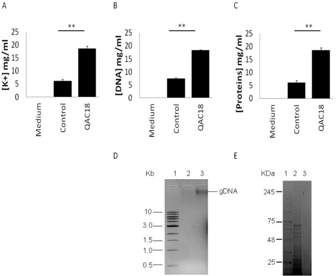
Figure 1.
Extrusion of cytoplasmic constituents of A. castellanii trophozoites to the external milieu after QAC treatment. [K+] (A), [DNA] (B) and [proteins] (C) released from 105 A. castellanii trophozoites after treatment without (control) and with QAC18 at 37.5 µg/ml for 24 h into spent medium and measured with the Atomic Absorption Spectroscopy (A), the Nanodrop at 260 nm (B) by Bradford assay (C) respectively were increased 3.6-fold, 4.1-fold and 15.1-fold respectively. Data is mean ± SD; n = 4 independent experiments performed in triplicates. **Student’s t-test showed significant difference of [DNA], [proteins] and [K+] in the spent media of control and QAC18-treated cells at p < 0.01. Extracellular DNA was visualised on 0.75% agarose gel (D), Key: Lane 1; DNA ladder, Lane 2; spent media on untreated trophozoites, Lane 3; spent media of QAC18-treated trophozoites and extracellular proteins visualised on SDS-PAGE (E). Key; Lane 1; protein ladder, Lane 2; spent media of QAC18-treated trophozoites, Lane 3; spent media on untreated trophozoites.

