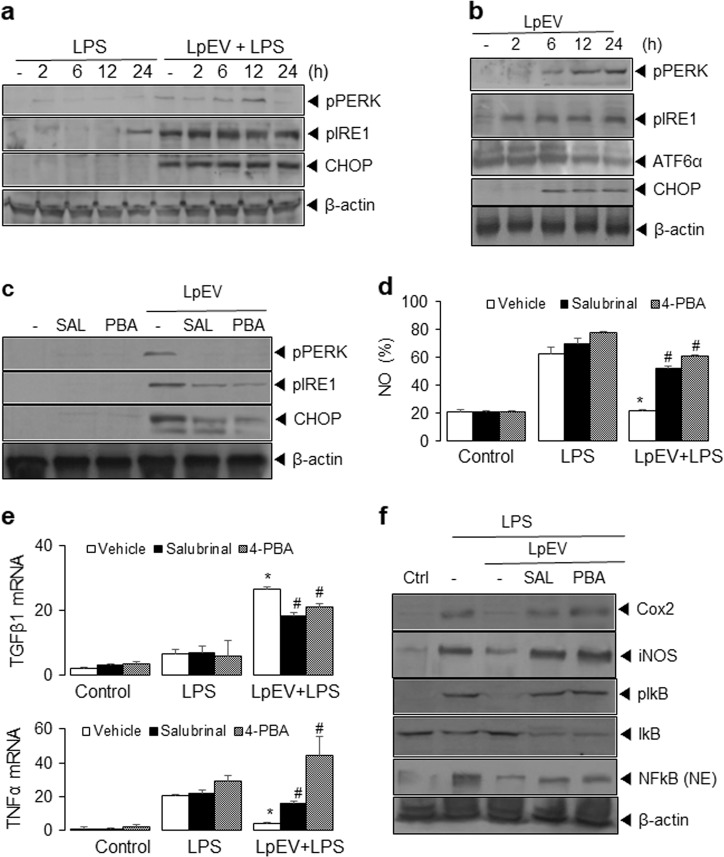
Fig. 6. Effect of LpEVs on ER stress-associated proteins in HT29 cells.
a HT29 cells were pretreated with vehicle or 500 ng/mL LpEVs and then treated with 1 mg/mL LPS for 0, 2, 6, 12, and 24 h. b HT29 cells were treated with 500 ng/mL LpEVs for 0, 2, 6, 12, and 24 h. a, b Cell lysates were resolved by SDS-PAGE and analyzed by western blot analysis with antibodies against CHOP, p-PERK, p-IRE1, ATF6α (p90), and β-actin. c HT29 cells were preincubated with 10 µM salubrinal or 5 mM 4-PBA for 1 h and treated with LpEVs for 12 h followed by LPS for 12 h. NO production was determined by a 2,3-diaminonaphthalene assay. d HT29 cells were preincubated with 10 µM salubrinal or 5 mM 4-PBA for 1 h, treated with LpEVs for 12 h, and then stimulated with LPS for 12 h. Cell lysates were analyzed by western blot analysis. e HT29 cells were preincubated with 10 µM salubrinal or 5 mM 4-PBA for 1 h, treated with LpEVs for 12 h and then stimulated with LPS for 0, 12, and 24 h. The mRNA levels of TNFα and TGFβ were then analyzed by qRT-PCR. f HT29 cells were preincubated with 10 µM salubrinal or 5 mM 4-PBA for 1 h, treated with LpEVs for 12 h, and then stimulated with LPS for 0 and 24 h. Cell lysates were analyzed by western blot analysis with antibodies against iNOS, pIκB, IκB, and NFκB. All data were plotted as the mean±standard deviation of at least three experiments. *P < 0.01 compared with LPS-treated cells without LpEVs. #P < 0.01 compared with LPS-treated cells with LpEVs.

