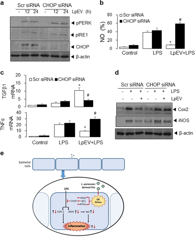
Fig. 7. Effect of LpEVs on CHOP expression in HT29 cells.
a HT29 cells were transfected with scrambled (Scr) control or CHOP siRNA for 24 h and then consecutively treated with LpEVs for 12 h and LPS for 0, 12, and 24 h. NO production was determined by a 2,3-diaminonaphthalene assay. b HT29 cells were transfected with scrambled (Scr) control or CHOP siRNA for 24 h and then treated with LpEVs for 0, 12, and 24 h. Cell lysates were resolved by SDS-PAGE and analyzed by western blot analysis. c, d HT29 cells were transfected with scrambled (Scr) control or CHOP siRNA for 24 h and then consecutively treated with LpEVs for 12 h and LPS for 24 h. c The mRNA levels of TNFα and TGFβ were then analyzed by qRT-PCR. d Cell lysates were analyzed by western blot analysis with antibodies against COX-2, iNOS, and β-actin. The results shown are representative of those obtained in more than three independent experiments. *P < 0.01 compared with LPS-treated cells without LpEVs. #P < 0.01 compared with LPS-treated cells with LpEVs. e The proposed model of LpEV-induced ER stress against LPS-induced inflammation in HT29 human colorectal cells. LPS induces COX-2, iNOS, and NFκB activation, resulting in NO production and inflammatory cytokine activation. LpEVs increase the ER stress response and the activation of PERK, IRE1, and ATF6α, leading to CHOP expression, which attenuates LPS-induced inflammatory responses.

