Figure 1.
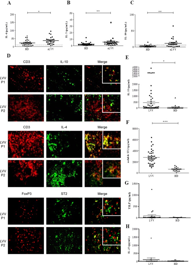
Expression of Th2 and anti-inflammatory cytokines in LVV. (A–C) PBMC of active LVV patients (aLVV) (n = 23) and HD (n = 27) were stimulated for 4 hours with PMA-ionomycine. Expression levels of interleukin 4 (IL-4), interleukin 5 (IL-5) and interleukin 10 (IL-10) measured in supernatant are represented. The levels of IL-4, IL-5 and IL-10 are higher in LVV patients as compared to HD [p = 0.01; p = 0.002 and p = 0.003, respectively]. We used a Mann Whitney test. These data are shown as the mean ± SEM. (D) Immunofluorescence analysis of inflammatory lesions from LVV patients reveals the expression of Th2 (here IL-4) and IL-10 cytokines partially by CD3 positive cells. Expression of ST2 by Treg (defined as FOXP3 cells) by immunofluorescence analysis. (E) Expression level of IL-33 in LVV (n = 44) patients serum was higher compared to HD (n = 18), *P < 0.05. (F) Mean level of soluble ST2 was increased in LVV patient sera (n = 38) as compared to HD (n = 17), ***P < 0.001. (G) Level of TSLP in LVV (n = 16) patients serum compared to HD (n = 9). (H) Level of IL-25 in LVV (n = 16) patients serum compared to HD (n = 9).

