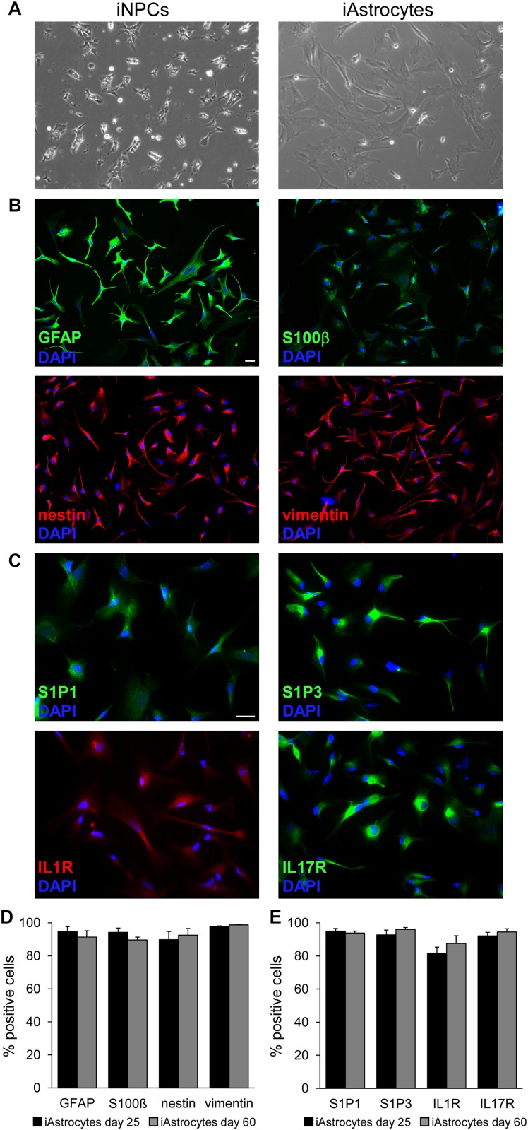
FIGURE 1.
Characterization of iPSC-derived astrocytes. (A) Phase contrast images showing human iNPC (left panel) and human iAstrocytes (right panel). (B,C) Representative immunofluorescence stainings for GFAP, S100β, nestin, vimentin (B) and S1P1, S1P3, IL1R and IL17R (C) in human iAstrocytes. DAPI was used for nuclear staining. (D,E) Percentage of cells positive for astrocyte markers at two timepoints during differentiation. Reported quantifications were performed on three different human iAstrocytes preparations from the same iNPC cell line. Bars represent SEM. Same observations were recorded in human iAstrocytes from a second iNPC cell line. Scale bar = 30 μm.

