Figure 2.
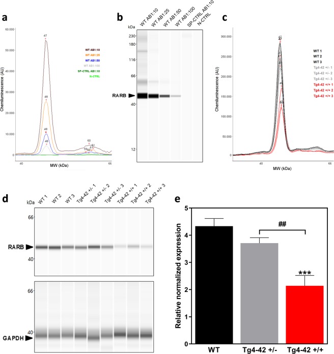
Simple Western protein analysis of the retinoic acid receptor beta in wild type (WT) and Tg4–42 + /+ mice. (a, b) Different antibody dilutions of a monoclonal retinoic acid receptor beta 2 antibody were tested with the Simple Western blot system to determine its binding capacity and optimal antibody concentration. A specificity control (SP-CTRL) and a negative control (N-CTRL) were used to detect unspecific binding of the column and cross reactions, respectively. (c, d) After protein extraction, striatal protein samples of three different WT, Tg4–42 + /− and Tg4–42 + /+ mice were measured by the Simple Western system. GAPDH was used as loading control. (e) Quantification of RARB protein of three wild type (WT), three hemizygous (Tg4–42 + /−) and three homozygous Tg4–42 (+/+) striatal samples as shown in (c, d) normalized to GAPDH. One-way ANOVA followed by Tukey’s multiple comparisons test (n = 3 per group. Mean + SEM. F (2, 6) = 43.4, R2 = 0.9354, Significances are labelled as followed: ***p < 0.001 against WT and ##p < 0.01 as indicated).

