Figure 1.

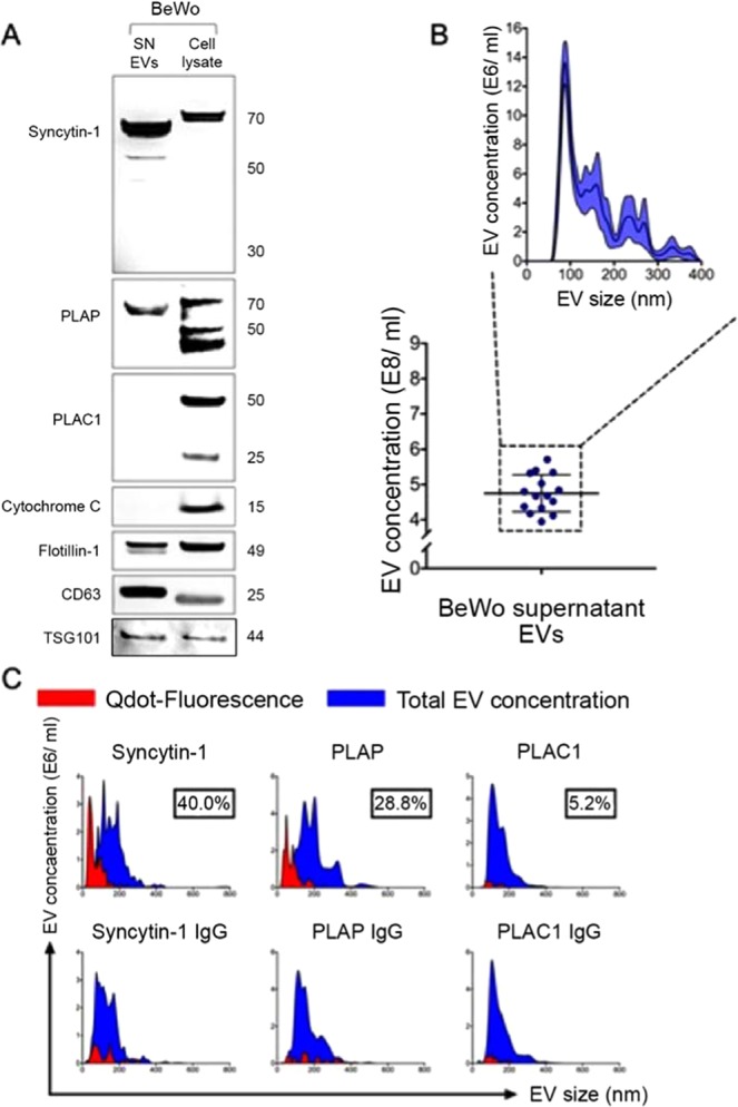
EVs released by BeWo cells express placenta specific protein markers. (A) Western blot analysis of EVs released into culture supernatant from BeWo cells. Supernatant EVs were positive for canonical exosomal markers CD63 and flotillin-1, suggesting that EVs isolated utilizing methodology detailed yielded nanoparticles. There was no contamination from cellular debris and apoptotic bodies in EV fractions (cytochrome C). BeWo EVs showed high expression of placental proteins syncytin-1 and PLAP, but not of PLAC-1. The blots were cropped from different gels. (B) Nanoparticle scatter analysis of BeWo EV preparations showed similar size distribution, with majority of EVs in the size range of 50–100 nm along with lower concentrations of microvesicles of larger sizes. (C) NanoSight nanoparticle detector analysis for surface expression of placental proteins is shown. Higher abundance of placenta-specific markers syncytin-1 and PLAP was noted compared to PLAC-1, which was not elevated compared to IgG isotype background fluorescence. EV subpopulations positive for syncytin-1, PLAP, and PLAC-1 (red) are shown in relation to the total EV pool (blue). Appropriate IgG isotypes (mouse IgG, rabbit IgG) were used as negative controls.
