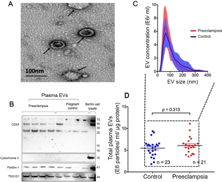
Figure 2.
EVs isolated from maternal circulation express canonical exosome markers. (A) A representative of Cryo EM image of plasma EVs isolated from normal pregnant individual (scale bar 100 nm). (B) EVs isolated utilizing methodology detailed yielded nanoparticles enriched in exosome markers CD63, TSG101 and flotillin-1 without contamination from cellular debris and apoptotic bodies (cytochrome C). The blots were cropped from different parts of the same gel. (C) Nanoparticle scatter analysis confirmed that majority of isolated EVs had size distribution consistent with exosomes (50–100 nm), and there was no difference in the size distribution of EVs between the Preeclampsia versus Control groups (p = 0.415). (D) Total plasma EV quantities in Preeclampsia versus Control groups were also similar (p = 0.313).

