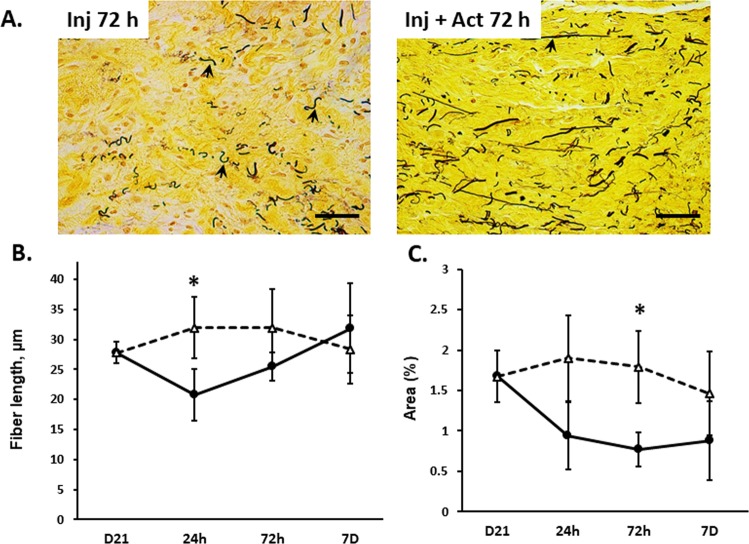
Figure 4.
Effect of injury on vaginal elastic fibers with injury ± actinonin. Microscopic images of vaginal muscularis on 3d postpartum in (A) injury + PBS (Inj 3d) and injury + actinonin (Inj + Act 3d). The 4–5 µm sections of full-thickness vaginal rings were stained with Hart’s stain. Quantitative analysis performed using a Nikon E1600 microscope with 40x objective and ImageJ software. Scale bars 50 µm. Graphical depiction represents (B) elastin fiber length (µm) and (C) percent area covered by elastic fibers in pregnant (D21) controls compared to postpartum time points for injury with (triangles) and without actinonin (solid circles). n = 4–5 in all groups, *P < 0.05 compared with injury alone time point, t-test.

