Figure 5.
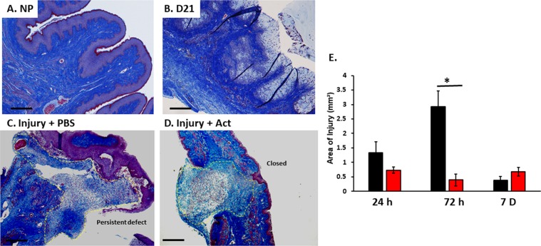
Representative histologic images of nonpregnant, day 21 and postpartum vaginal wall after injury. Microscopic images of vaginal muscularis and epithelium in nonpregnant (NP) (A), day 21 (B), 3d after delivery in injury + PBS (C) and injury + actinonin (D). The 4–5 µm sections of full-thickness vaginal rings were stained with Masson trichrome. Quantitative analysis of area of injury (outlined with yellow dashed line. Vaginal rings in the actinonin treated specimen (D) are intact with statistically smaller area of injury. Injury + PBS specimens (C) maintain persistent defects at injury site. (E) Area of injury (mm²) for postpartum injured animals treated with PBS (black) or actinonin (red). PBS, phosphate buffered saline; Act, actinonin. 4X magnification, Scale bar, 500 µm.

