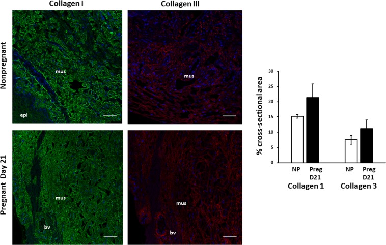
Figure 8.
Immunolocalization of Collagen Types I and III in the vaginal wall of pregnant and nonpregnant rats. Immunofluorescence of Collagen I (LEFT) and III (RIGHT) in uninjured rat vaginal wall in nonpregnant virginal rats (top row) and pregnant rats on day 21 of gestation (bottom row). Sections of full-thickness vaginal rings (4–5 µm) were labeled with primary antibodies to collagen I and III. Green = collagen I, Red = Collagen III, Blue = DAPI. Increased ratio of collagen I/III noted in each specimen. Increased content of both collagen subtypes noted in pregnant specimens. Scale bars 50 µm. Graphical depiction (Right) of collagen area between nonpregnant controls (NP) and pregnant (Preg D21) animals. mus, vaginal muscularis; epi, epithelium; bv, blood vessel.

