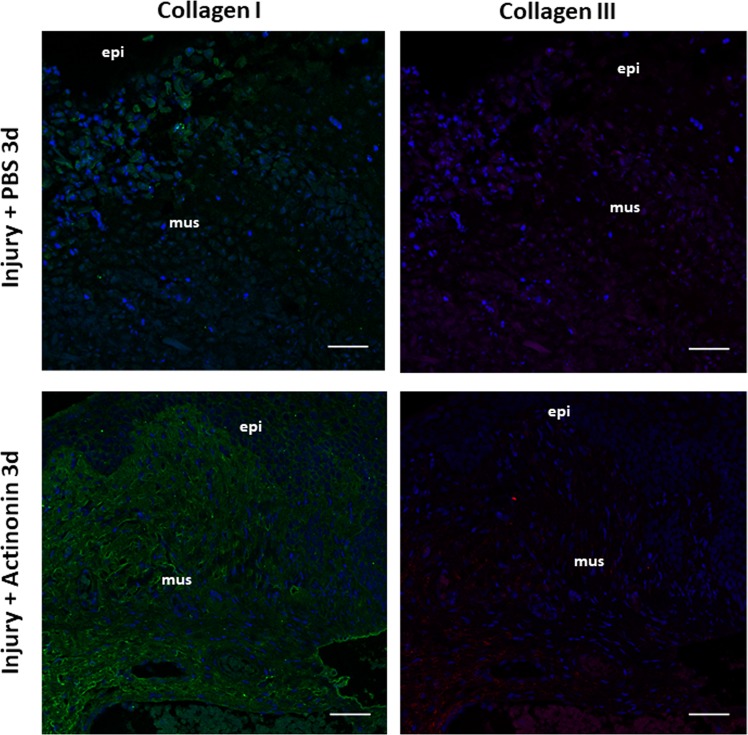
Figure 9.
Immunolocalization of Collagen Types I and III in the vaginal wall after obstetrical injury. Immunofluorescence of Collagen I (1st column) and Collagen III (2nd column) in rat vaginal wall at site of injury 3 days after delivery in injury+ PBS (top row) and Injury+ actinonin (bottom row). Green = collagen I, Red = Collagen III, Blue = DAPI. Scale bars 50 µm. PBS, phosphate buffered saline; Act, actinonin; mus, vaginal muscularis; epi, epithelium.

