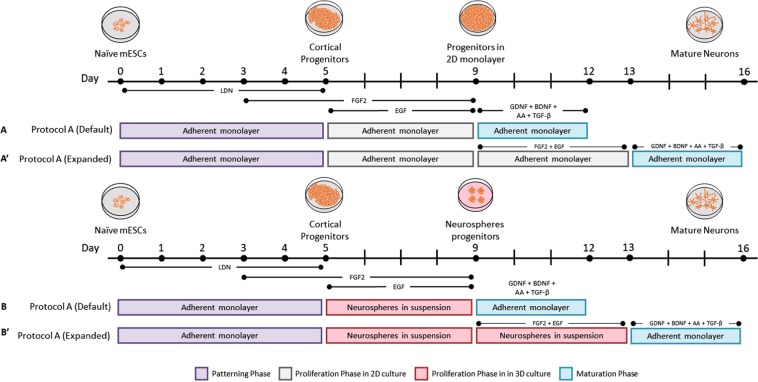
Figure 1.
Schematic overview depicting the experimental design of the two neural induction protocols, highlighting the morphogens and small molecules employed in the current study. (A,A′) In the first protocol, mESCs were first patterned for 5 days, proliferated for 4 days -or (A′) passaged to be cultured for another 4 days, and lastly subjected to maturation for 3 days all of which were conducted in a 2-dimensional microenvironment. (B,B′) The second protocol was carried out in a similar fashion as the 2D protocol except that mESCs were cultured in a 3-dimensional microenvironment during the proliferation phase allowing them to grow in sphere-like structures (neurospheres).

