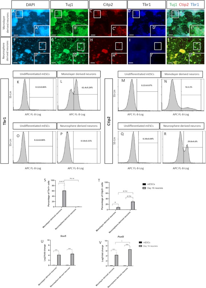
Figure 4.
A panel of representative immunostaining images of 16 day old mature neurons derived from (upper) 2D monolayer and (lower) 3D neurosphere cultures. Monolayer-derived neurons labelled with (A,A′) DAPI, confirming their maturation by expressing (B,B′) Tuj1 and further confirming their cortical linage being positive for (D,D′) Tbr1 and with modest expression for (C,C′) Ctip2. (E,E′) Merged images. Neurosphere-derived neurons labelled with (F,F′) DAPI, showing their positivity for (G,G′) Tuj1 and (H,H′) Ctip2 but not (I,I′) Tbr1. (J,J′) Merged images. Flow cytometry comparative analysis of (K–N) Tbr1 and (O–R) Ctip2 expressions between monolayer- and neurosphere-derived neurons with their corresponding undifferentiated counterparts. A comparative analysis of the positively stained monolayer- and neurosphere-derived neurons for (S) Tbr1 and (T) Ctip2 comparing to their undifferentiated counterparts. Log-2-fold change in the transcriptional expression of (U) Sox5 and (V) Fezf2 supporting the deep layer cortical identity. Data represents mean ± SEM, n = 5, Students t-test *p < 0.05, ***p < 0.001. Scale bars = 100 um.

