Figure 1.
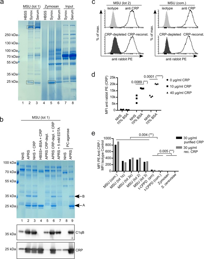
CRP binds to MSU crystals. (a) Synovial fluid or serum from a patient with pseudogout was incubated with MSU crystals (lot 2) or zymosan for 45 min at 37 °C. Unbound proteins were washed away and bound proteins were eluted and subjected to SDS-PAGE and visualized by coomassie. Bands excised for mass spectrometric analyses are indicated (rectangles). (b) Normal human serum (NHS), acute phase reaction serum (APRS; CRP around 100 µg/ml) or HBSS (always containing Ca2+) with or without depletion of CRP or addition of purified CRP to 100 µg/ml were incubated with MSU crystals or phosphorylcholine-agarose (PC-agarose) for 45 min at 37 °C. Bound proteins were eluted and subjected to SDS-PAGE and visualized by coomassie. In addition, the same samples were analyzed by Western blot analysis using CRP antibody (lower panel) or C1qB antibody (middle panel). (c) Human serum (CRP = 10.4 µg/ml) was left untreated or CRP was depleted with PC-agarose or was depleted and then reconstituted with 10 µg/ml purified CRP. All three sera were incubated with two preparations of MSU crystals (lot 1 and a commercial preparation (com.)). CRP was stained with CRP antibody and anti-rabbit-PE and analyzed using a flow cytometer. (d) Three different preparations of MSU crystals (lot 1, lot 2 and a commercial preparation) were incubated with either pool serum (NHS) (CRP < 0.3 µg/ml) or 10% BSA in HBSS, both with purified CRP added to the indicated concentrations. Binding of CRP to the crystals was analyzed as in c. Median fluorescent intensity (MFI) is shown. (e) Four different preparations of MSU crystals (one commercial (com) and three self-made (untreated or sonicated (s)), two preparations of t-CPPD (commercial (com.) and self-made (sm)) and two preparations of S. Cerevisiae (zymosan and heat-inactivated yeast) were incubated with NHS (CRP < 0.3 µg/ml) with either 30 µg/ml purified CRP or 30 µg/ml recombinant (rec.) CRP added. Binding of CRP to the crystals and fungal particles was analyzed as in c. MFI of staining with CRP antibodies was divided by MFI of isotype controls. Uncropped images of gels and Western blot are shown in Fig. S3.

