Figure 2.
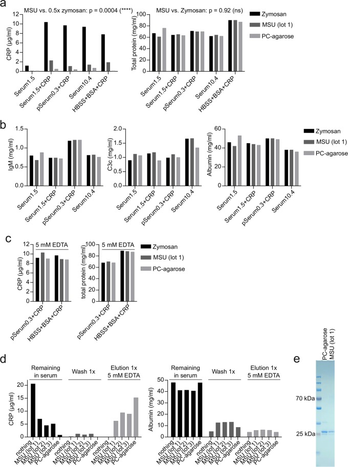
MSU crystals specifically purify CRP (a) 200 µl of serum of a single donor with 1.5 µg/ml CRP (Serum1.5) with or without addition of 10 µg/ml purified CRP, a pool serum with 0.3 µg/ml CRP (pSerum0.3) with 10 µg/ml purified CRP added, a single donor serum with 10.4 µg/ml CRP (Serum10.4) and HBSS 10%BSA with 10 µg/ml purified CRP added, were incubated with 3 mg zymosan, 5 mg MSU (lot 1) or 35 µl PC-agarose for 45 min at 37 °C. Samples were centrifuged and the supernatants were analyzed for CRP and total protein concentration. Using a one-sample t-test, the p-value of MSU samples compared to 50% of CRP concentration of the corresponding zymosan sample was calculated. For the difference of total protein in zymosan or MSU treated samples a paired t-test was used. (b) The concentration of IgM, C3c and albumin was analyzed in samples from a by turbidimetry. (c) For the indicated CRP-containing solutions the experiment was repeated in the presence of 5 mM EDTA and the supernatants were analyzed for CRP and total protein concentration. (d) 200 µl of pool serum containing 20 µg/ml purified CRP was incubated with nothing, three distinct preparations of MSU (5 mg each) or 35 µl PC-agarose for 45 min at 37 °C, washed 1x with HBSS for 5 min and then eluted with 5 mM EDTA in HBSS. Supernatants of each step were analyzed for CRP and albumin concentration by turbidimetry. (e) 100 µl of a serum containing 30 µg/ml CRP was incubated with 9 mg MSU or 35 µl PC-agarose for 45 min at 37 °C. MSU/PC-agarose was washed 5x in HBSS, and CRP was eluted by HBSS + 5 mM EDTA. Eluted proteins were applied to SDS-PAGE and proteins were visualized by coomassie staining. Uncropped image of the gel is shown in Fig. S3 (right gel). Each experiment was repeated at least once with similar results.

