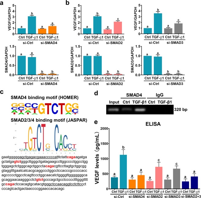
Fig. 4. SMAD signaling pathways are involved in TGF-β1-induced VEGF expression and secretion.
a SVOG cells were transfected with 50 nM control siRNA (si-Ctrl) or SMAD4 siRNA (si-SMAD4), and after 48 h they were treated with 5 ng/mL TGF-β1 for 6 h. VEGF and SMAD4 mRNA levels were examined by RT-qPCR. b SVOG cells were transfected with 50 nM control siRNA (si-Ctrl), SMAD2 siRNA (si-SMAD2) or SMAD3 siRNA (si-SMAD3), and after 48 h they were treated with 5 ng/mL TGF-β1 for 6 h. VEGF, SMAD2, and SMAD3 mRNA levels were examined by RT-qPCR. c Photo of SMAD-binding motifs in the HOMER database and JASPAR database (upper panel). The SMAD-binding site in the human VEGF promoter is highlighted in red. The primers for the ChIP assay are underlined (lower panel). d SVOG cells were treated with 5 ng/mL TGF-β1 for 1 h before being subjected to ChIP analysis. Anti-SMAD4 or IgG antibodies were used to immunoprecipitate DNA-containing complexes. Subsequent PCR was performed with primers that were complementary to the VEGF promoter region and contained the SMAD-binding site. The PCR products were resolved by electrophoresis on a 1% agarose gel and were visualized by ethidium bromide staining. e SVOG cells were transfected with 50 nM control siRNA (si-Ctrl), SMAD4 siRNA (si-SMAD4), SMAD2 siRNA (si-SMAD2), SMAD3 siRNA (si-SMAD3) or SMAD2 + SMAD3 siRNAs (si-SMAD2 + 3), and after 48 h they were treated with 5 ng/mL TGF-β1 for 24 h. The protein levels of VEGF in culture media were examined by ELISA. The results are expressed as the mean ± SEM of at least three independent experiments. The values without a common letter are significantly different (p < 0.05).

