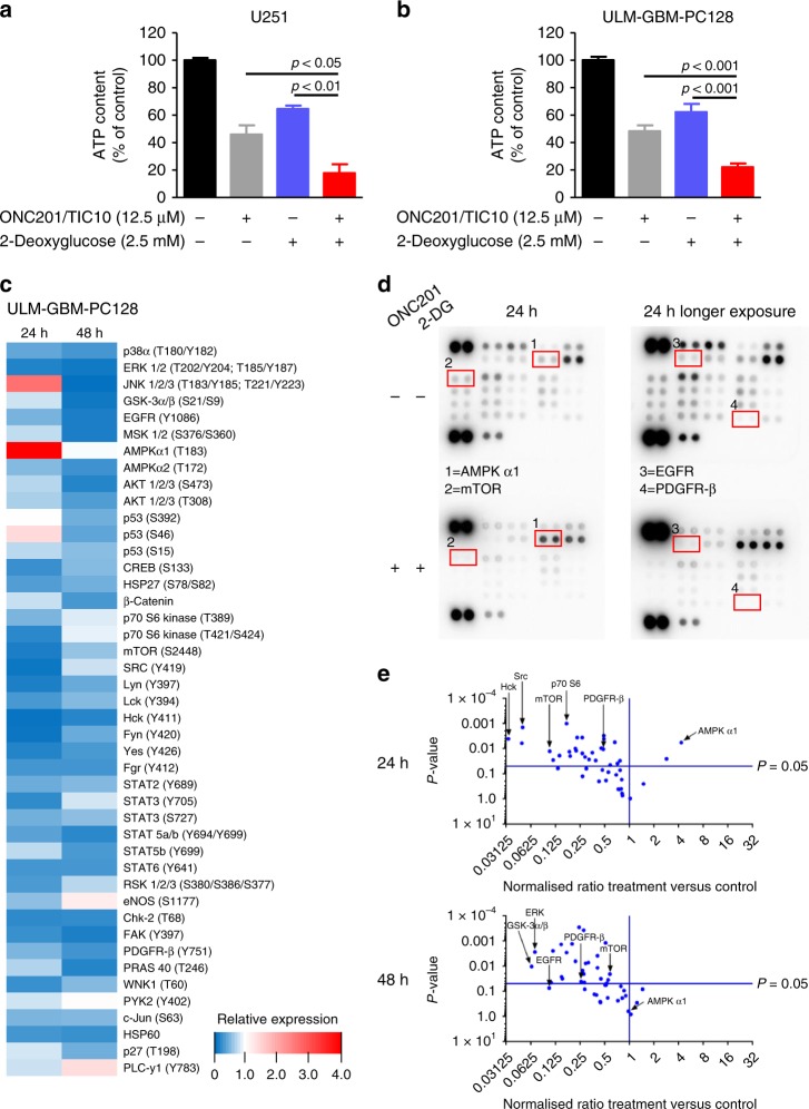
Fig. 3. Treatment with ONC201/TIC10 and 2-Deoxyglucose causes energy deprivation and a hypo-phosphorylative state.
a, b U251 (a) and ULM-GBM-PC128 (b) cells were treated for 72 h as indicated. CellTiter-Glo® assays were performed to determine ATP levels. Columns, mean; bars, SD; n = 3. c, ULM-GBM-PC128 cells were treated for 24 h or 48 h with 10 µM ONC201/TIC10 and 1.25 mM 2-Deoxyglucose combined or the respective solvent prior to collecting the lysates. After incubation of the respective lysates with the membranes of the human phospho-kinase array kit (R&D Systems, ARY003b) bound phospho-proteins were detected according to the manufacturer’s instructions. Each membrane contains kinase-specific and reference antibodies spotted in duplicate. A key to identify the coordinates of each protein spotted on the array is available on the R&D System’s website (http://www.rndsystems.com/pdf/ARY003b.pdf). Quantitative analysis using the Bio-1D software (Vilber Lourmat) was performed and the data were first normalised to the respective reference spot and second to the respective treatment control. The relative phospho-kinase expression (ON201/TIC10 plus 2-Deoxyglucose versus control) is displayed in a heatmap. d Representative autoradiography of the human phospho-kinase array described in c after 5 min/10 min exposure. e Volcano plots comparing Log2 fold change of phosphorylated phospho-proteins for the combination treatment versus control after 24 h and 48 h.

