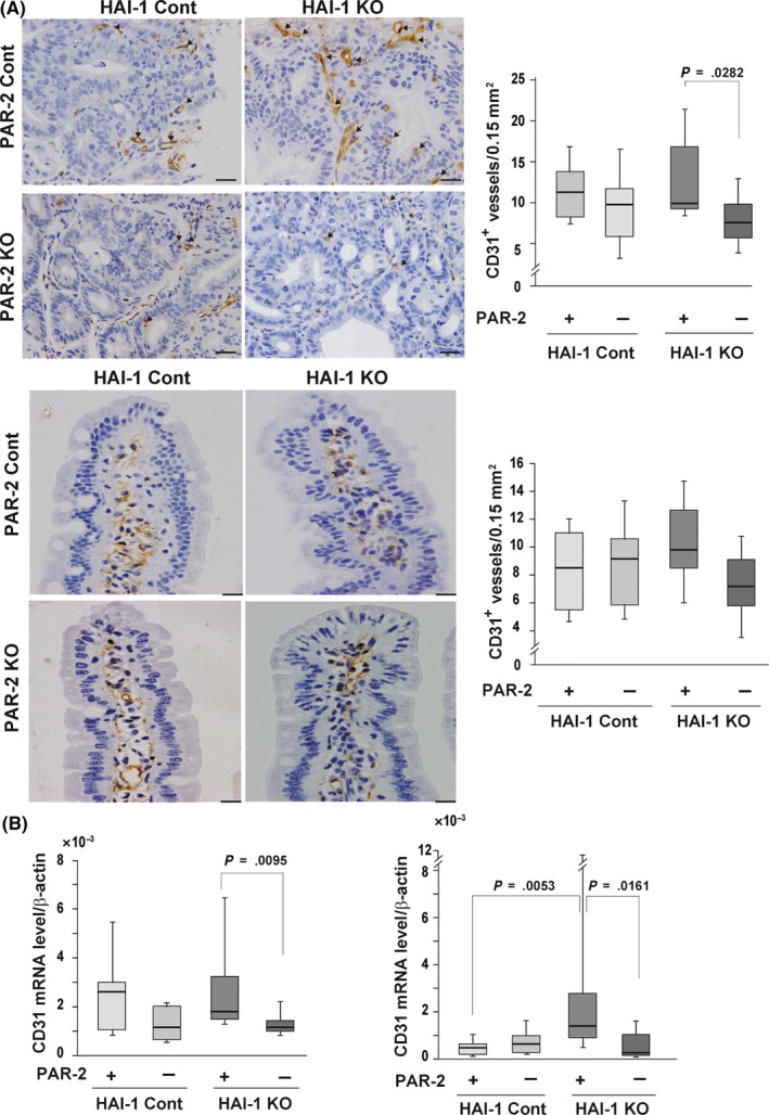
Figure 5.

Effect of protease‐activated receptor‐2 (PAR‐2) deficiency on capillary density. A, Immunohistochemical analyses of CD31. Representative photographs of CD31+ vessels (arrows) (left panel), quantitation of CD31+ vessels (right panel) of tumor tissues (upper panel), and nontumor mucosa tissues (lower panel) from Spint1LoxP/LoxP/Apc Min/+ mice (hepatocyte growth factor activator inhibitor‐1 [HAI‐1] control [cont] + PAR‐2 cont) (n = 7), Spint1LoxP/LoxP/F2rl1 −/−/Apc Min/+ mice (HAI‐1 cont + PAR‐2 knockout [KO]) (n = 6), Spint1LoxP/LoxP/Villin‐Cre/Apc Min/+ mice (HAI‐1 KO + PAR‐2 cont) (n = 5), and Spint1LoxP/LoxP/Villin‐Cre/F2rl1 −/−/Apc Min/+ mice (HAI‐1 KO + PAR‐2 KO) (n = 5). Bar = 20 μm. B, Quantitative RT‐PCR for Pecam1 (CD31) mRNA expression in tumor (left panel) and nontumor mucosa (right panel) tissues. Box and whiskers indicate the interquartile range and sample maxima and minima, respectively. Median is indicated by a bold line
