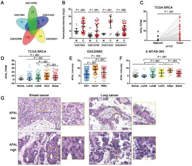
Figure 1.
Expression of Aurora A/PLK1–associated long noncoding RNAs (lncRNAs) (APAL) in cancer tissues. A) Venn diagram of differentially expressed lncRNAs in five breast cancer datasets with fold changes greater than 1.5 (P < .01) as analyzed by two-sided independent sample t test. B) APAL expression in breast cancer datasets as analyzed by independent-sample two-sided t test. C) APAL expression in the tumors and normal tissues of paired TCGA breast cancer data (BRCA) data as analyzed by two-sided paired sample t test. D–F) APAL expression in different subtypes of breast cancer in The Cancer Genome Atlas (TCGA) BRCA (n = 813), GSE20685 (n = 327), and E-MTAB-365 (n = 426) datasets. G) APAL expression in representative paraffin-embedded tissues of breast cancer (n = 199) and lung cancer tissues (n = 64), as detected by in situ hybridization (ISH). ISH signals are detected as blue-purple staining with nuclei counterstained by nuclear fast red. Scale bar = 50 μm. See also Supplementary Figure 1 (available online).

