
Figure EV2. Anti‐GA antibodies block the non‐cell‐autonomous effects of poly‐GA on TDP‐43 in a co‐culture assay.
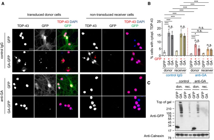
Primary hippocampal neurons were transduced with GFP or GA175‐GFP (DIV4 + 4) and treated with IgG control and anti‐GA (5F2) antibody.
- Confocal imaging revealed that anti‐GA antibody treatment reduces Poly‐GA‐induced cytoplasmic mislocalization of TDP‐43 in hippocampal neurons. White and red arrows show cells with cytoplasmic TDP‐43 in GFP‐positive and GFP‐negative cells, respectively. Scale bar denotes 20 μm.
- Automated quantification of cells with cytoplasmic TDP‐43 in GFP or GA175‐GFP‐transduced cells. Cells with and without GFP signal were analyzed separately (indicated by +/−). As in Fig 1C, GFP‐negative donor and GFP‐positive receiver cells were excluded due to high transduction and low transmission rate of GFP. n = 4 biological replicates. Scatter plot with bar graphs of mean ± SD. One‐way ANOVA with Tukey's multiple comparisons test. *P < 0.05, and ***P < 0.001.
- Immunoblotting shows reduced poly‐GA expression upon anti‐GA antibody treatment.